Cardiac Unit
The Cardiac Unit is a non-invasive Cardiac Laboratory that provides care and diagnostic tests/procedures to patients with a history of or suspected 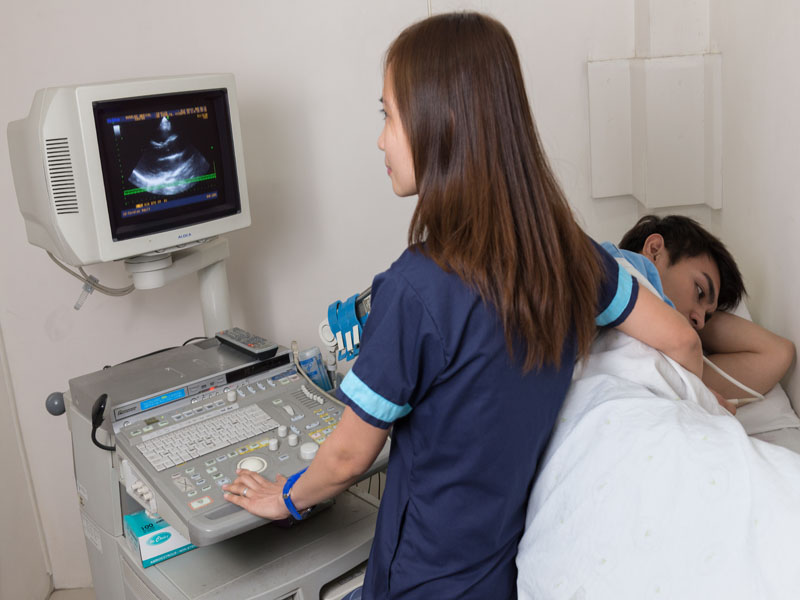 Cardiovascular Disease, (i.e. chest pain, history of collapse, palpitations, suspected Coronary Artery Disease or an irregular heart rhythm) non- invasive diagnostic tests and procedures that will effectively aid and complement our consultant Physician in their critical mission to devise adequate and sufficient management and treatment under their care.
Angeles Medical Center, Inc. Cardiac Unit include a team of Echo cardiographers who are well-trained and have expertise in and 2D-echocardiography technologists who are also trained, customer-oriented and efficient. All staff members received training in Basic Life Support and Advanced Cardiac Life Support. We intend to consistently deliver reliable, timely and accurate diagnostic results in the service and support of our clients, physicians and patients. Our unit’s vision is to become the preferred and most respected diagnostic center in the Central Luzon region and in other community that will serve.
Diagnostic tests provided/offered:
Two-Dimensional Echocardiogram (2D-ECHO)
Often referred to as a cardiac echo or simply an echo. A sonogram of the heart. High frequency sound waves are used to create a Two-dimensional and Doppler ultrasound to create images of the heart. The test is performed with a device, called transducer, placed on the chest.
It is capable of displaying a cross sectional “slice” of the beating heart, including chambers, valves and major blood vessels that exit from the left and right part of the heart.
Is a safe and painless diagnostic procedure that uses high frequency sound waves to take moving pictures of the heart. The sound waves are directed at the heart from a small, hand held transducer which sends and receives signals that appear on the screen as a moving image of the heart.
Electrocardiogram (ECG/EKG)
Safe, non-invasive procedure, used to measure the heart’s electrical conduction system. It records the changes in magnitude and direction of the electrical activity of the heart. The electrodes placed in standard positions on the body todetect the electric current generated by depolarization and repolarization of the atria and ventricles. The voltages generated are amplified and recorded on ECG paper as waves and complexes.
Treadmill Stress Test
An exercise test, gathers information about how the heart works during physical 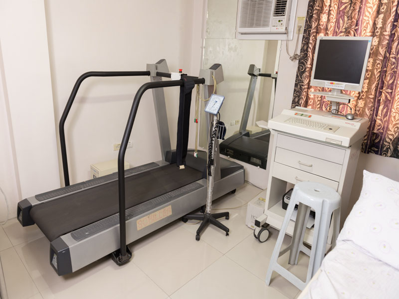 activity. It involves walking or running on a treadmill while the heart rhythm, blood pressure and breathing are monitored. It is used to guide treatment for patient diagnosed with a heart condition and recommended for patient with symptoms of Coronary Artery Disease.
Holter Monitor (24hr)
A Holter monitor is a machine that continuously records the heart’s rhythm. The monitor is worn for 24 hours during normal activity.
How the Test is performed
Electrodes (small conducting patches) are stuck onto your chest. These are attached by wires to a small recording monitor. You carry the Holter monitor in a pocket or pouch worn around your neck or waist. The monitor runs on batteries.
While you wear the monitor, it records your heart’s electrical activity. Keep a diary of what activities you do while wearing the monitor, and how you feel. After 24 hours, you will return the monitor to Heart Station’s reception desk. The doctor will look at the records and see if there have been any abnormal heart rhythms.
Holter Heart Monitor
It is very important that you accurately record your symptoms and activities so the doctor can match them with your Holter monitor findings. Electrodes must be firmly attached to the chest so the machine gets an accurate recording of the heart’s activity. While wearing the device, avoid: Electric blankets, High-voltage areas, Magnets, and metal detectors.
Continue your normal activities while wearing the monitor. You may be asked to exercise while being monitored if your symptoms have occurred in the past while you were exercising.
How to prepare for the Test
You do not need to prepare for the test. Your doctor will start the monitor. You’ll be told how to replace the electrodes if they fall off or get loose. Tell your doctor if you are allergic to any tape or other adhesives. Make sure you shower or bathe before you start the test. Yow will not be able to do so while you are wearing a Holter monitor.
How the test will Feel
This is a painless test. However, some people may need to have their chest shaved so the electrodes can stick. You must keep the monitor close to your body. This may make it hard for you to sleep.
Why the Test is Performed
Holter monitoring is used to determine how the heart responds to normal activity. The monitor may also be used after a heart attack, to diagnose heart rhythm problems, and when starting a new heart medicine.
It may be used to diagnose atrial fibrillation or flutter, multifocal atrial tachycardia, reasons for fainting, slow heart rate (bradycardia), and ventricular tachycardia.
Normal Results
Normal variations in heart rate occur with activities. A normal result is no significant changes in heart rhythms or pattern.
What Abnormal Results Mean
Abnormal results may include various arrhythmias. Changes may mean that the heart is not getting enough oxygen. The monitor may also detect conduction block, a condition in which the atrial electrical activity is either delayed or does not continue into the ventricles of the heart.
Risks
There are no risks associated with the test. However, you should be sure not to let the monitor get wet.
Alternative Name
Ambulatory Electrocardiography
Diana Ocno
[email protected]
(045) 888 7150/ (045) 322 4632/ (045) 9414 local 596
AMCI- West Wing Building, Ground Floor; Monday to Saturday 8:00am – 5:00pm
NOTE: Electrocardiogram (ECG/EKG) procedure is available at the Emergency Department when the Cadiac Unit is closed.